 |
|
||||||||||
|
|
 |
|
|||||||||
|
|
|
 |
|
||||||||
|
|
|
||||||||||
|
|
|
||||||||||
|
|
|
||||||||||
|
|
|
||||||||||
|
|
|
||||||||||
|
2008, Vol. 3 No. 2, Article 28
Isolation of Candida spp. from Mastitic cows and Milkers M. A. Tarfarosh* and S. K. Purohit
*Corresponding Author;
Institute of Animal Health and Biological Products, Srinagar
ABSTRACT Fifty six cases of bovine clinical mastitis were screened for presence of Candida spp. Of these Candida was isolated from milk of four cases (7.14%). The organism was also isolated from 80% of milkers of such animals. KEY WORDS Candida spp., mastitis, thrush, milkers. INTRODUCTION
Candida is the most common species isolated from cases of mycotic mastitis in bovines (Radostitis, 1995). The organism and its spores have the ability to survive pasteurization; as such it assumes public health significance and has been indicated in causation of
thrush in humans (Schmitt, 1971).
MATERIAL AND METHODS
Milk samples from 56 clinical cases of bovine mastitis were aseptically collected at the college clinic and other veterinary hospitals in Bikaner city. About 20ml of milk was collected from each affected animal after discarding first few strips. The samples were rushed to laboratory under cold conditions and incubated at 37°C for 24 hours and there after streaked on Saborauds glucose agar plates. The plates were incubated at 37°C and examined for growth at 24, 48 and 72 hours and at biweekly intervals for 4 weeks after which the plates showing no growth were considered negative.
RESULTS AND DISCUSSION
Out of the 56 cows suffering from clinical mastitis only 4 (7.14%) were found to harbour
Candida infection. These findings are comparable with observations of Bansal et al (1991) and Singh et al (1992) who reported incidence of
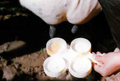
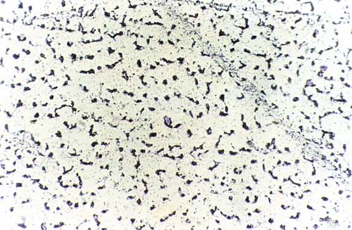
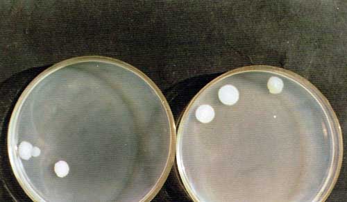
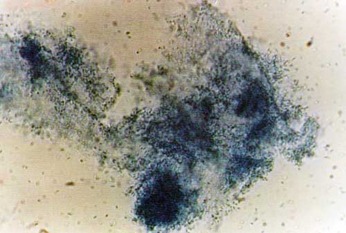
Candida mastitis as 8.51% and 5.71% respectively. Typical Candida colonies (fig 4) were observed on Saborauds agar plates. The microscopic examination of teased mounts revealed pseudo-hyphae, clusters of budding cells, blastospores and chlamydospores (fig 3 & 5). Clinical examination of the affected animals revealed inflammation, tenderness and hardening of the udder with secretion of watery milk containing yellowish clots (fig 1).
REFERENCES
Fig. 1: Inflammation and hardening of udder with discoloration of milk
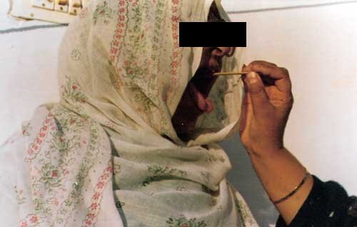
Fig. 2: Swabs were taken from the buccal mucosa of the milkers of affected cows
Fig. 3: Microscopic slides of the growths obtained on Saborauds glucose agar medium
Fig. 4: Candida colonies on Saborauds agar plates
Fig. 5: Another slide prepared from teased mounts
|
|
||||||||||
|
|
|||||||||||
|
|
|||||||||||
|
|
|||||||||||
|
|
|||||||||||
|
Copyright © Vet Scan 2005- All Right Reserved with
VetScan |
Home | e-Learning |Resources | Alumni | Forum | Picture blog | Disclaimer |
|
|||||||||
|
powered by eMedia Services |
|
||||||||||
|
|
|
|
|
|
|
|
|
|
|
|
|