 |
|
|||||||||||||||||||||
|
|
 |
|
||||||||||||||||||||
|
|
|
 |
|
|||||||||||||||||||
|
|
|
|||||||||||||||||||||
|
|
|
|||||||||||||||||||||
|
|
|
|||||||||||||||||||||
|
|
|
|||||||||||||||||||||
|
|
|
|||||||||||||||||||||
|
2009, Vol. 4 No. 2, Article 45
Experimental Osteo-fluorosis in Goats of Jammu and Kashmir Vinay Kant*1, P.K.Verma2, N. K. Pankaj2 and R. Raina3
1M.V.Sc. Scholar; 2Junior Scientist; 3Professor and Head;
Division of Pharmacology and Toxicology,
*Corresponding Author; e-mail address: [email protected]
ABSTRACT Fluorine intoxication is an important global public health concern in humans and animals. In present study, sodium fluoride alone and with aluminium sulphate (ameliorative agent) was administered orally daily for 30 days in healthy goats of group 1 and 2 respectively to access the radiographic changes in the forelimb and efficacy of aluminium sulphate as an ameliorative agent. The radiographic changes that were observed in both the groups after 30 days exposure include an increase in overall density of the bony cortex. No other significant change s in the bone and or the joint of any of the animals were detected. KEY WORDS Aluminium sulphate, goats, radiographic changes, sodium fluoride, subacute toxicity. INTRODUCTION
Fluoride toxicity is one of the serious health problems in many parts of the globe and is an important public health concern in humans and animals. In India, toxicity of fluoride in man and animals is very common and about 20 states including Jammu & Kashmir (J&K) have been identified as significantly affected states in India ( Thakuria, 2007). In Jammu & Kashmir state excess fluorine level in soil and water is present in some belts of district Doda, Kathua and Rajouri. Many people of Ghat area in Doda district are reported to be suffering from dental fluorosis. The water in this area has the level of fluoride is over 1.2 ppm which exceeds the national standards for fluoride in drinking water (1 ppm). The dental fluorosis is spreading to the adjacent rural areas of Doda (Majeed, 2007). In the J&K state, goats are back bone of livestock economy and main source of livelihood of Gujjars and Bakerwals community.
MATERIALS AND METHODS
Eight healthy cross bred goats 1.5 -2 years of age weighing 25-30 kg were procured from local farmers of R.S. Pura, Jammu. They were acclimatized for two weeks in the divisional animal shed under standard conditions before the commencement of experiment. The animals were maintained on ad libitum feed and water. The experimental protocol was approved by institutional animal ethics committee. The animals were divided into two groups of 4 each. Goats of group 1 were used to study the effect of subacute exposure of fluoride on radiographs of forelimb, in which sodium fluoride (NaF, SD Fine- Chem. Ltd.) alone was administered orally at the dose rate of 20 mg/kg body wt. (providing 9 mg/kg b. wt. fluorine) daily for 30 days and goats of group 2 were used to study the efficacy of aluminium sulphate as an ameliorative agent on the effect of subacute intoxication of fluoride on radiographic changes, in which same dose of NaF along with aluminium sulphate [SD Fine- Chem. Ltd.] at the dose rate of 150 mg/kg b.wt. was administered orally daily for 30 days. Aluminium sulphate was administered 30 minutes before the administration of NaF. Both the salts were dissolved separately in 100 ml of distilled water and gavazed to the animals daily between 9.00 to 10.00 a.m. All the animals were weighed weekly and doses of NaF and Al2(SO4)3 were corrected accordingly.
RESULTS AND DISCUSSION
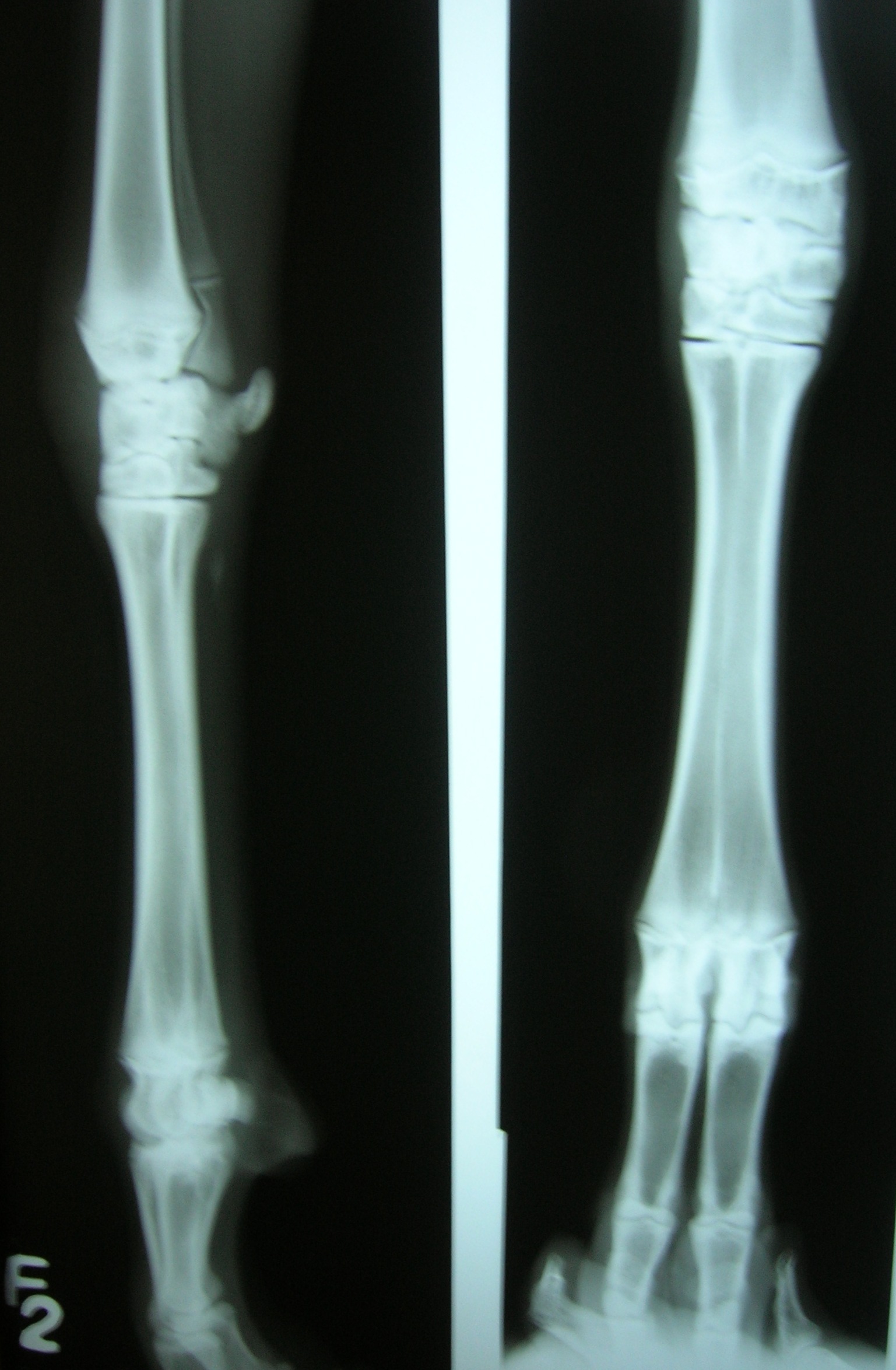
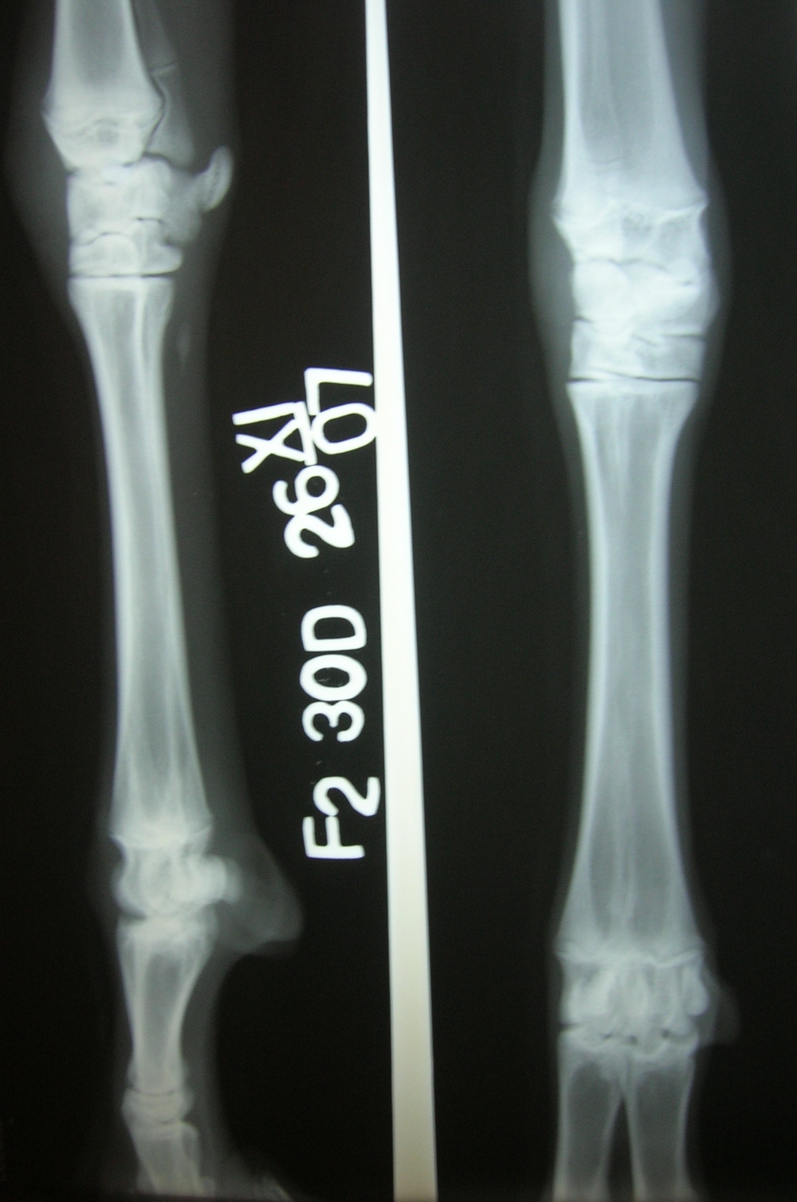
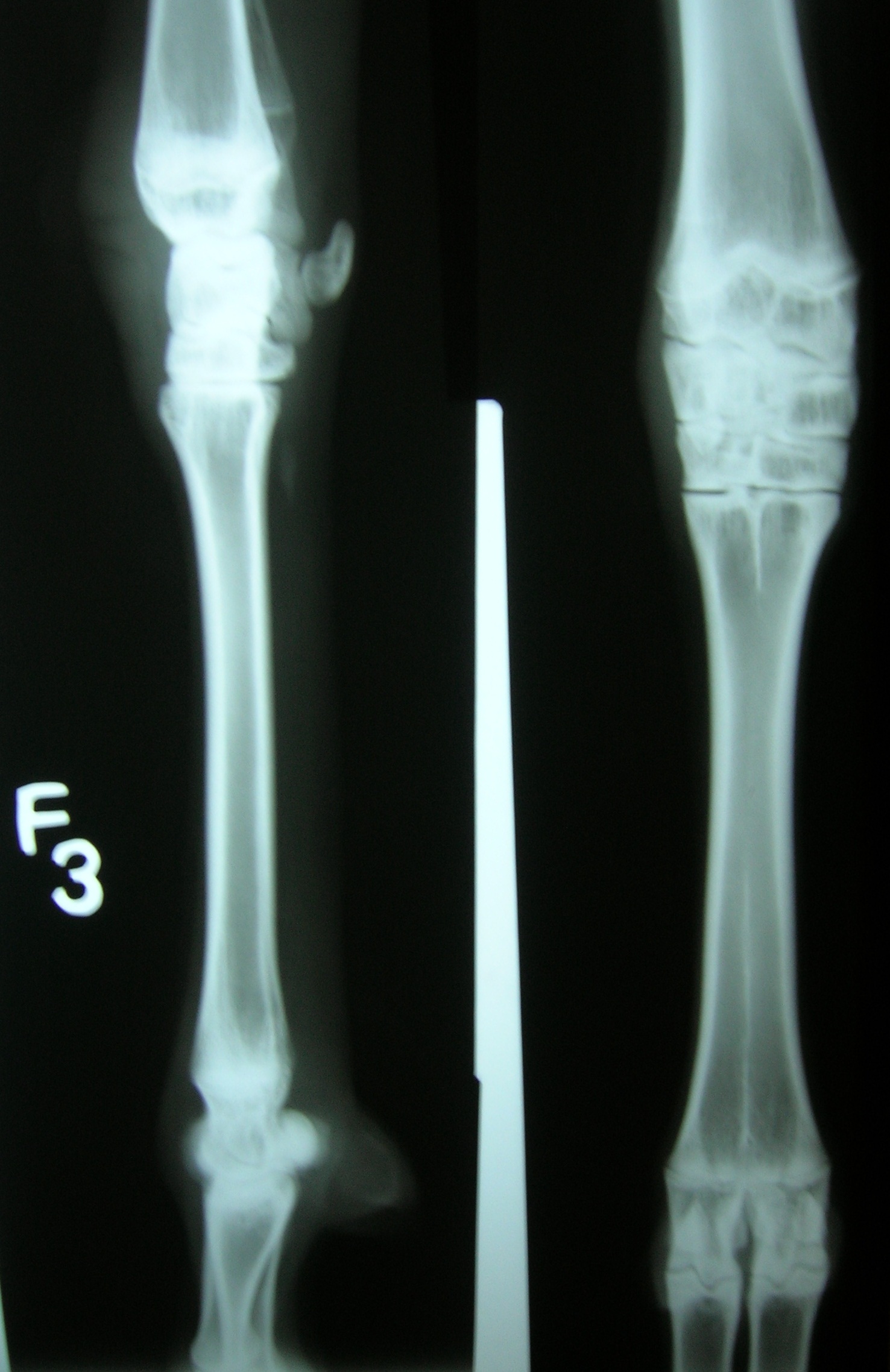
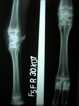
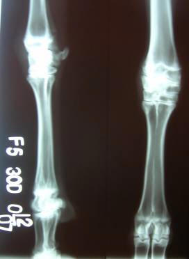
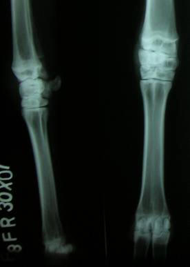
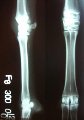
The radiographs of metacarpal of right forearm of two goats of group 1 and 2 are presented in Figure 1 and 2, respectively. The radiographic changes that were observed after 30 days exposure of sodium fluoride include only an increase in overall density of the bony cortex. No other significant changes in the bone and or the joint of any of the animals were detected. These less prominent changes in the bones and the joints may be due to short time fluoride exposure and goats are more resistant to fluoride toxicity among the ruminants. Similar types of changes in the radiographs were also observed in Group 2. So, the use of aluminium sulphate as an ameliorative salt had no beneficial effects on the changes produced in the bones. During the experiments, goats showed decreased movement, symptoms of lameness and muscle weakness. They sat and stayed at one place during grazing.
REFERENCES
Fig 1: Radiographs of right forelimb, with lateral and anterio-posterior view,
Fig 2: Radiographs of right forelimb, with lateral and anterio-posterior view,
|
|
|||||||||||||||||||||
|
|
||||||||||||||||||||||
|
|
||||||||||||||||||||||
|
|
||||||||||||||||||||||
|
|
||||||||||||||||||||||
|
Copyright © Vet Scan 2005- AAll Right Reserved with
VetScan |
Home | e-Learning |Resources | Alumni | Forum | Picture blog | Disclaimer |
|
||||||||||||||||||||
|
powered by eMedia Services |
|
|||||||||||||||||||||
|
|
|
|
|
|
|
|
|
|
|
|
||||||||||||