 |
|
|||||||||||||||||||||||||||||||||||||||||||||
|
|
 |
|
||||||||||||||||||||||||||||||||||||||||||||
|
|
|
 |
|
|||||||||||||||||||||||||||||||||||||||||||
|
|
|
|||||||||||||||||||||||||||||||||||||||||||||
|
|
|
|||||||||||||||||||||||||||||||||||||||||||||
|
|
|
|||||||||||||||||||||||||||||||||||||||||||||
|
|
|
|||||||||||||||||||||||||||||||||||||||||||||
|
|
|
|||||||||||||||||||||||||||||||||||||||||||||
|
2009, Vol. 4 No. 2, Article 40
Studies on Management of Dental Root Abscess and Related Osteomyelitis in Domestic Animals A. S. Bigham1*, S. N. Dehghani2, Z. Shafiei3 and H. Khosropanah4
Department of Veterinary Surgery and Radiology, Faculty of Veterinary Medicine, Shahre-kord University, Shahre-kord, Iran.
Department Of Veterinary Surgery & Radiology, School of Veterinary Medicine, Shiraz University, Shiraz, Iran.
Department Of Periodentology, Shiraz University Of Medical Science, Shiraz, Iran.
*Corresponding Author; e-mail address: [email protected]
ABSTRACT Seven animals of different species suffering from dental problems were referred to the surgical unit of the schools. Six of them had upper or lower jaw osteomyelitis along with dental root abscesses and one had dental fracture. All these animals were managed successfully by extra-oral and intra-oral extraction of the involved teeth. KEY WORDS Dental extraction, domestic animals, root abscess, osteomyelitis. INTRODUCTION Periodontal disease is the most common infectious disease in humans and animals. 75% to 80% of cats and dogs over 2 years of age exhibit signs of periodontal infection (5). In animals the upper premolar teeth (generally the fourth premolar tooth) whose roots are located near the eye socket are mostly affected. These animals are generally presented with swollen eye and fistula. Treatment includes extraction of the tooth and flushing of the infected site with antibacterial solution. A drain is required in severely infected cases (6). Present study reports successful management of dental root abscess along with lower or upper jaw osteomyelitis, in seven animals of different species. MATERIALS AND METHODS
The study was conducted on Seven animals of different species having different dental problem ( table 1). Extraction/repulsion of the diseased teeth was performed after radiographic examinations.
The
premedication in dog ,horse and cat involved tranquilization with acepromazine acetate (0.2 mg/kg, IM) followed by Anaesthesia with ketamine hydrochloride (5-10mg/kg, IV) and diazepam (0.2 mg/kg, IV) and maintained with inhalant anaesthesia (2% Halothane), whereas sheep and cow was sedated with xylazine (0.02 mg/kg, IM) and local dental anaesthesia using 2% lidocaine was achieved prior to extraction of diseased teeth.
Affected teeth in cat, dog, sheep and cow were extracted in the following three steps:
RESULTS and discussions Disappearance of facial wounds and satisfactory alveolar cavity granulation was recorded within 10-20 days post antibiotic treatment (Table 2) in all cases. The horse has normal host defenses that work to maintain the integrity of the tissues that support the tooth. A disruption of the defense mechanisms results in an opportunistic infection (7). In small animal practice tooth extraction is usually performed through the oral approach under general anesthesia (2). In horses the tooth extraction technique is known as repulsion. It requires an osteotomy or trephination to expose the roots of the diseased tooth. Extraction is achieved by driving/repelling the tooth into the mouth with a dental punch (8). After extraction, flushing and gentle debridement of the alveolus removes loose fragments, debris and infected apical tissues (9). This can be done using an intra-oral or extra-oral approach. In all species, the extraction site is generally sutured as this enhances wound healing, prevents contamination from food particles, stops hemorrhage effectively and reduces postoperative pain (3,9). This procedure is contra-indicated, however, in cases of active infection where one should allow drainage of infected debris or discharges (10). Providing extra-oral drainage to infected areas after tooth extraction procedures (approached through cortical bone) in lagomorphs and horses, allows primary closure of the alveolus (1). Postoperative antibiotic administration is indicated in the presence of an active infection; severe periodontal disease or osteomyelitis (4,1,10,11). REFERENCES
Table 2: Post operative
antibiotic therapy in different animals.
Fig 1:Ventero- dorsal (1) and lateral view (2) radiographs of maxillary osteomyelitis and 4th premolar dental root abscess 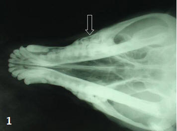 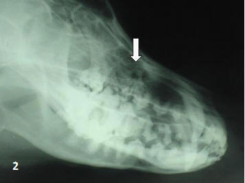
|
|
|||||||||||||||||||||||||||||||||||||||||||||
|
|
||||||||||||||||||||||||||||||||||||||||||||||
|
|
||||||||||||||||||||||||||||||||||||||||||||||
|
|
||||||||||||||||||||||||||||||||||||||||||||||
|
|
||||||||||||||||||||||||||||||||||||||||||||||
|
Copyright © Vet Scan 2005- All Right Reserved with
VetScan |
Home | e-Learning |Resources | Alumni | Forum | Picture blog | Disclaimer |
|
||||||||||||||||||||||||||||||||||||||||||||
|
powered by eMedia Services |
|
|||||||||||||||||||||||||||||||||||||||||||||
|
|
|
|
|
|
|
|
|
|
|
|
||||||||||||||||||||||||||||||||||||