|


2010, Vol. 5 No. 2, Article 67
A Histo-anatomical Study of the Trachea of
Central Asian Tortoise
(Testudo horsfieldii) in Iran
M. Shadkhast*, H. R. Shabazkia1, A. S. Bigham2, S. E. Shariati, T. Mahmoudi
Department of Anatomical and Histological Science of Basic Science,
Faculty of Veterinary Medicine, Sharekord University, Shahrekord, Iran
1Department of Biochemistry and Pharmacology,
Faculty of Veterinary Medicine, Sharekord University, Shahrekord, Iran
2Departement of Veterinary Surgery and Radiology,
Faculty of Veterinary Medicine, Sharekord University, Shahrekord, Iran
*Corresponding Author;
e-mail address: [email protected]
ABSTRACT
Histology of the tracheal epithelium has been studied in various species including reptiles
like the common lizard (Lacerta agilis). However, meager information is available on this aspect in case of Asian tortoise (Testudo horsefieldii). A study was therefore conducted on (n=10) central Asian tortoise (five male and five female) to describe the microscopic structure of the tracheal wall. The observations revealed that trachea constituted mainly of complete cartilage rings lined by a respiratory epithelium (ciliated, basal and mucous cells) with variable morphology. The organ was short and divided into paired bronchi which remained separated along the sides of the neck.
KEY
WORDS
Morphological study, tortoise, trachea.
INTRODUCTION
The structure of the epithelium of trachea has been widely studied in guinea-pig (Dalen, 1983), mouse
( Jeffery and Reid, 1975; Pack et al, 1981), rabbit (Plopper et al, 1984) and different reptilian species (Pastor et
al, 1987; Tesik, 1987 & 1992). However, the structure and tracheal morphology of the central Asian tortoise has received little attention. The information available on this aspect is very scanty in reptiles and mainly refers to lizards and turtle (Tesik loc. cit. and Pastor et
al loc.cit.). The present investigations was therefore undertaken to describe
the microscopic structure of tracheal wall of the central Asian tortoise.
MATERIALS AND METHODS
The study was conducted on the trachea of (n=10) adult central Asian tortoise (five males and five females) at educational hospital of
Faculty of the Veterinary Medicine, Sharekord University, Shahrekord, Iran.
Tissue samples from the trachea were fixed by immersion in 10% buffered formalin.
Samples were routinely processed and embedded in paraffin. Five micron thick sections were stained with haematoxylin and eosin, Van Giesson and Periodic acid Schiff (PAS).
RESULTS AND DISCUSSION
The observation revealed that a well
defined trachea extended from end of pharynx nearly to the heart, where it bifurcated. The framework of thin tubular structure consisted of 60±1.2 superimposed complete rings of hyaline cartilage covered with loose connective tissue.
Average length of the organ was 82 ±1.9mm (mean±SD) with inner lining consisting of a layer of epithelial tissue.
Light microscopic examination showed the trachea to be lined with a pseudostratified
columnar epithelium that consisted of basal, ciliated and mucous
cells. The basal cells were located in the basal portion of the
epithelium and processes were occasionally observed passing towards
the lumen. These cells were present in abundance and differentiated clearly.
The basal cells arise by multiple divisions similar to germinal cells of the epidermis
(Blenkinsopp, 1967). Apparently the change of basal cell into the definitive type of the epithelial element occurs through an intermediate stage, represented by the intermediary cell. Presumably the whole process is completed so rapidly that the occurrence of intermediary cells in the epithelium becomes rare. Unlike basal cells, the intermediary cells are considerably less uniform. Their cytoplasm varies in density containing no
secretory granules. In Testudo horsefieldii, the goblet and granular cells arising from intermediary cells retain the typical morphological traits that characterize them as specific separate species. In birds and mammals it is possible to encounter cells bearing concurrently some traits of both granular and goblet element. Ciliogenesis in intermediary cells
of rats was observed by Jeffery and Reid (1975).
During the present investigation
tracheal epithelium showed variations in thickness and stratification depending on the area, especially in the cartilaginous and epithelial part from the larynx to the extra pulmonary bronchi. However, no differences were observed between the cartilaginous and the membranous zones. These observations were not however in agreement with finding of Tesik (1984 & 1992) who reported
abundant presence of granular cells in the tracheas of Lacerta vivipara and
Lacerta agilis; and also found ciliated, granular and goblet cells frequently in the tracheal epithelium of most of the reptiles. Their mutual numerical proportions varied with the order or family of the reptiles examined. The incidence of occurrence of goblet and granular cells was governed by the law of reciprocity. In the present study few lymphocytes and plasma cells were observed in the lamina propria
of tracheal bronchus whereas these cells are found in abundance in
mammals (Dalen, 1983; Jeffery and Reid 1975; Pack et al 1981). The
presence of well defined cartilaginous structures forming complete
circles in most portions of the trachea, which gradually became segmented in its
caudal region, indicates their importance in the maintenance of patency to air.
The trachea of Testudo horsefieldii was well defined, slightly different in structure from the trachea of reptiles studied so far and
resembling the trachea of mammals except for presence of complete hyaline cartilage rings.
REFERENCES
-
Dalen H: (1983); An ultrastructural study of the tracheal epithelium of the guinea-pig with special reference to the ciliary structure. Journal of Anatomy;136(Pt 1):47.
-
Jeffery PK, Reid L(1975): New observations of rat airway epithelium: a quantitative and electron microscopic study. Journal of Anatomy;120(Pt 2):295.
-
Pack RJ, Al-Ugaily LH, Morris G(1981): The cells of the tracheobronchial epithelium of the mouse: a quantitative light and electron microscope study. Journal of Anatomy;132(Pt 1):71.
-
Krouse R (1922). Mikroskopische Anatomie der Wirbeltiere. II. Vogel und Reptilien. Berlin und Leipzig: Vereinigung
wissenschaftlicher Verlag a Co
-
Tesik I(1984): The ultrastructure of the tracheal epithelium in European common lizard (Lacerta agilis L.) and in sand lizard (Lacerta vivipara Jacq.). Anatomischer Anzeiger;155(1-5):329.
-
Tesik I(1992): Less Common Cell Types in the Tracheal Epithelium of Reptiles. ACTA VETERINARIA BRNO;61:17
-
Pastor LM, Ballesta J, Hernandez F, Perez-Tomas R, Zuasti A, Ferrer C: A (1987)Microscopic study of the tracheal epithelium of Testudo graeca and Pseudemys scripta elegans. Journal of Anatomy;153:171.
-
Blenkinsopp WK(1967): Proliferation of respiratory tract epithelium in the rat. Exp Cell Res;46:144-54.
-
Kuehne B, Junqueira LCU(2000;): Histology of the trachea and lung of Siphonops annulatus (Amphibia, Gymnophiona). Revista Brasileira de Biologia 60:167-72.
-
Kennedy AR, Desrosiers A, Terzaghi M, Little JB(1978): Morphometric and histological analysis of the lungs of Syrian golden hamsters. Journal of Anatomy;125(Pt 3):527
-
McCarthy C, Reid L(1964): Acid mucopolysaccharide in the bronchial tree in the mouse and rat (sialomucin and sulphate). Experimental Physiology 1964;49(1):81.
-
Plopper CG, St George JA, Nishio SJ, Etchison JR, Nettesheim P(1984): Carbohydrate cytochemistry of tracheobronchial airway epithelium of the rabbit. Journal of Histochemistry and Cytochemistry;32(2):209.
-
Spicer SS(1965): Diamine methods for differentiating mucosubstances histochemically. Journal of Histochemistry and Cytochemistry;13(3):211.
FIGURES
Fig.1-Dorsal view of Testudo horsefieldii
(Central Asian Tortoise)
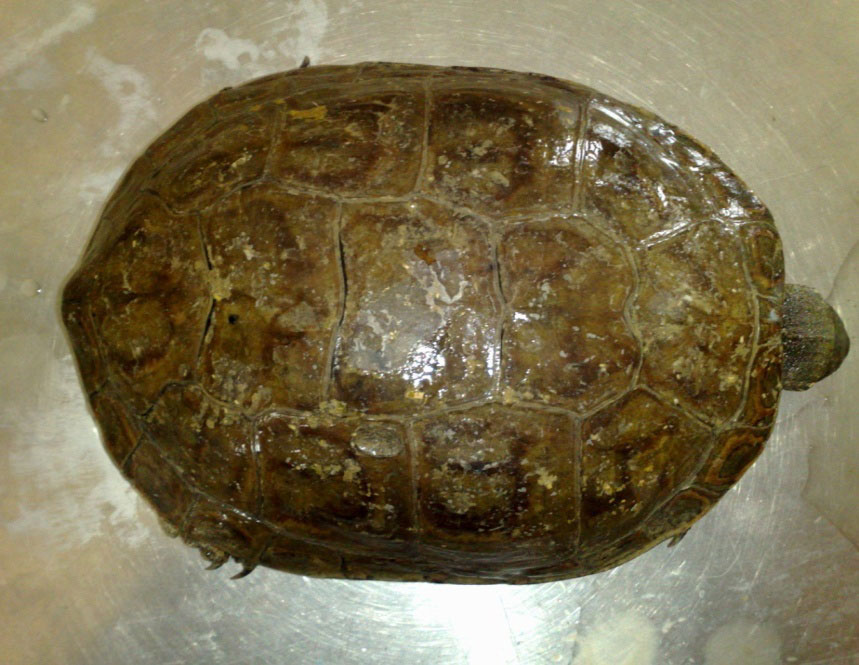
Fig. 2. Bifurcation of bronchials from trachea (white arrow)
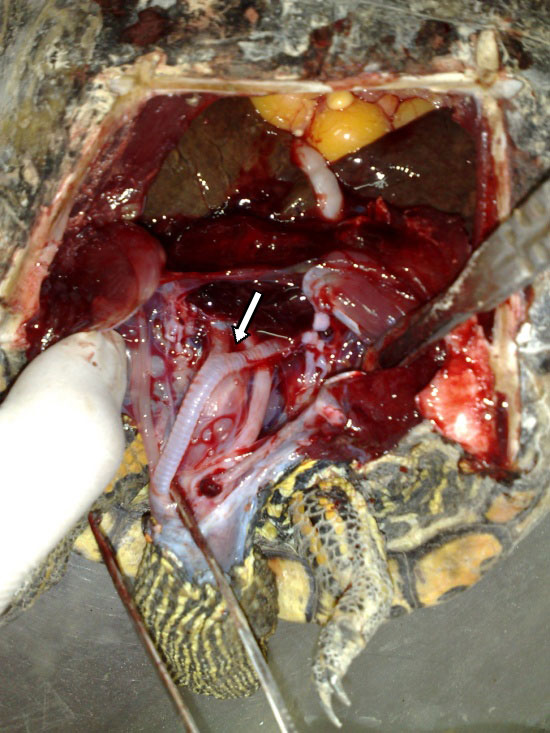
Fig. 3. Transvers section of the trachea. Epithelial lining separated from the cartilage by loose connective tissue (Van Giessen 4X)
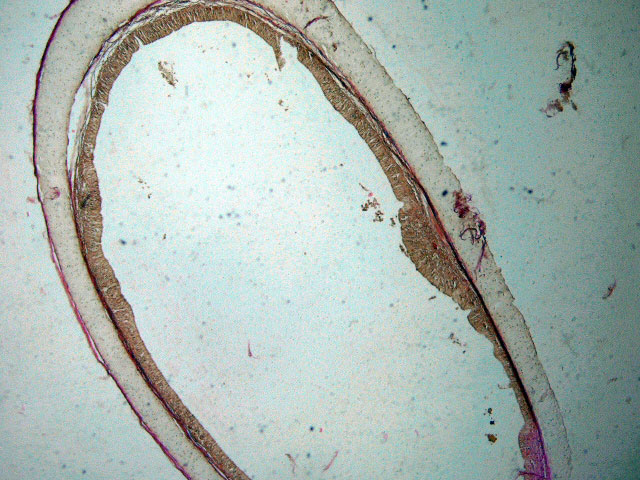
Fig. 4. Tracheal epithelium showing. Basal cell, ciliated and mucous cell (Van Giessen, 10X)
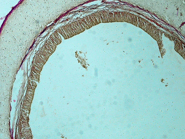
Fig. 5. Tracheal epithelium showing the goblet cells (white arrow), ciliated cells (yellow arrow) and basal cells (black arrow) (Van Giessen, 40X)
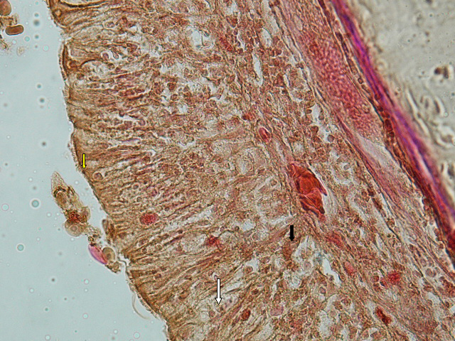
Fig. 6. Basal cell of tracheal epithelium - note the high vascularity of the connective tissue (Van Giessen, 40X)
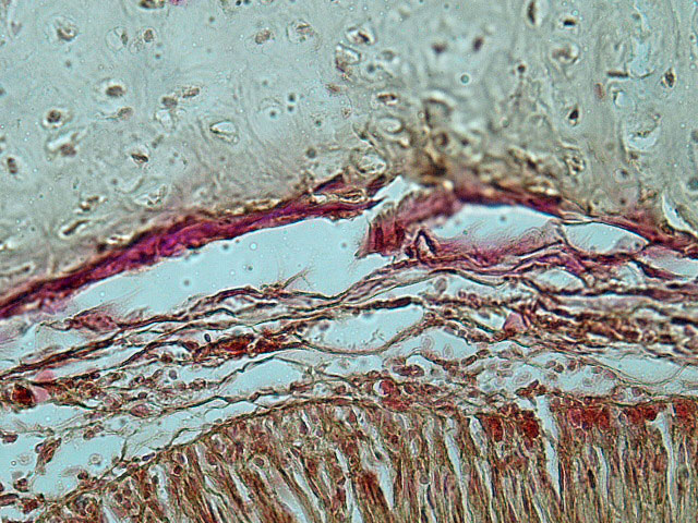
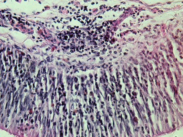
Fig. 7. The laminal propria of trachea showing abundant lymphocyte and plasma cells (PAS, 40X)
|
 |