 |
|
||||||||||
|
|
 |
|
|||||||||
|
|
|
 |
|
||||||||
|
|
|
||||||||||
|
|
|
||||||||||
|
|
|
||||||||||
|
|
|
||||||||||
|
|
|
||||||||||
|
2010, Vol. 5 No. 2, Article 72
Echinococcus infestation in lung of Nilgai
Geeta Devi Leishangthem1, Aparajita Choudhury2, Khangembam Victoria Chanu3* and Gautam Patra4
1Department of Pathology,
*Corresponding Author; e-mail address: [email protected]
ABSTRACT The present communication places on record an accidental finding ” hydatidosis” in a Nilgai that was under treatment for injury to hind limbs but succumbed subsequently to the wound infection. Postmortem examination revealed a single cyst protruding on the right anterior lobe of the lung. The cystic fluid stained with Gentian violet showed numerous hydatid sand of Echinococcus granulosus. Histopathological observations of the affected lung tissue and cyst wall were suggestive of hydatidosis. KEY WORDS Echinococcosis, histopathology, lung, Nilgai. INTRODUCTION
Echinococcosis or Hydatid disease is a zoonotic infection caused by cystic larval stage of tapeworm
Echinococcus granulosus. It is worldwide in distribution. Foci of hydatid disease also exist in India with highest prevalence in Andhra Pradesh and Tamil Nadu (11). In man the disease is characterized by a slow but steady progression of the parasitic lesions and a high mortality rate in untreated patients (1). The metacestode stage proliferates in the intermediate and aberrant hosts, predominantly in the liver by exogenous budding and by invading surrounding tissues comparable to a malignant tumour (2).
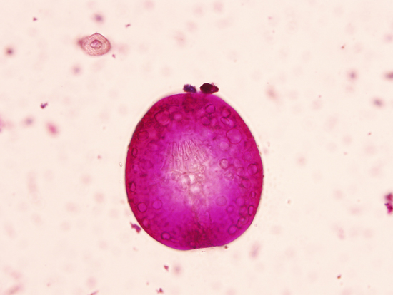
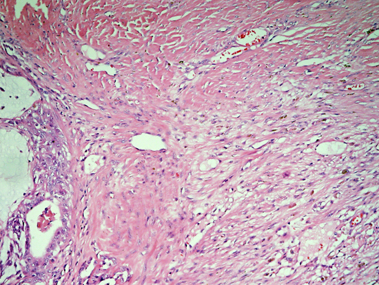
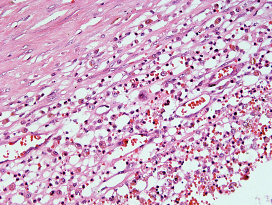
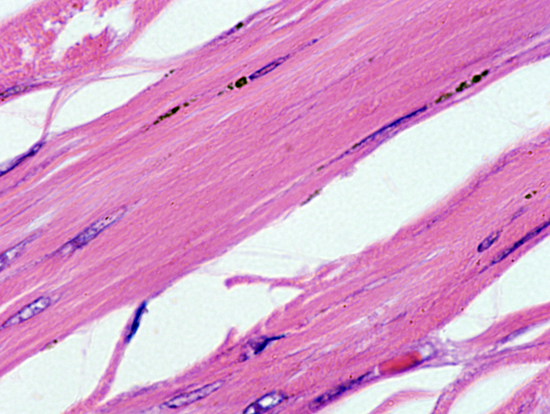
MATERIALS AND METHODS A female Nilgai (Boselaphus tragocamelus) was brought to the Veterinary Hospital, Society for Prevention of Cruelty to Animal (SPCA), Noida with injury to the hind limbs but it subsequently succumbed to wound infection. Post mortem examination revealed presence of a single cyst in the lung. The cystic fluid was collected and centrifuged at 1000 rpm for 10 min. The sediment was then stained with 3% Gentian violet. The lung was preserved in 10% buffered formalin and processed for routine histopathological examination. Following the routine procedure, the lung tissues were embedded in paraffin and sections of 5mm thickness were cut. The sections were then stained with hematoxylin and eosin and examined under microscope (10). RESULTS On post mortem examination, a single cyst measuring about 3x2.5x3 cm was found protruding on the right anterior lobe of the lung. The cyst contained about 15 ml of proteinaceous fluid. Upon drainage of fluid, the inner surface of the cyst wall showed a folded appearance and displayed numerous white colored granular structures. The fluid stained with Gentian violet revealed numerous hydatid sand of Echinococcus granulosus (Fig. 1). Histopathological examination of the lung tissue exhibited extensive tissue reaction characterized by various degrees of cellular infiltration and fibrosis along with numerous brood capsules attached to the cystic membrane (Fig. 2). The bronchial epithelium was hyperplastic and peri-bronchiolar blood vessels were engorged with blood. Necrotic material and many giant cells, surrounded by an outer layer comprising of macrophages, lymphocytes and a few numbers of epitheloid histocytes, were seen (Fig. 3). Outer wall of the cyst showed typical echinococcal hyalinous laminated membrane (Fig. 4). DISCUSSION
In the natural cycle, dogs and other canids are typical definitive hosts and a wide range of mammals, including sheep, cattle, pig, buffalo, goat, camel, horse and man, can serve as the intermediate host by
harboring the metacestode stage (12,13,14). The infection of carnivores with immature or mature intestinal stages of
E. granulosus does not cause morbidity, whereas the invasion of various organs (mainly liver and lungs) of intermediate or aberrant hosts by metacestode can cause severe and even fatal disease (hydatidosis). Man is an accidental intermediate host, as entry of the larval forms into humans represents an end stage in its life cycle. The outcome of infection in livestock and humans is cyst development in the liver, lungs, or other organ systems. The lungs and liver appear to be the sites of predilection (13).
ACKNOWLEDGEMENTS The authors are grateful to Chief, SPCA, Noida for providing the samples and Dr. Amit Kumar Dinda, Additional Professor, Department of Pathology, AIIMS, New Delhi for providing the facilities for histopathological studies. REFERENCES
FIGURES
Fig. 2 & 3: Histopathological sections of lung showing extensive tissue reaction along with inflammatory cells infiltrate around the necrotic areas
Fig. 4: Cyst wall showing laminated membrane
|
|
||||||||||
|
|
|||||||||||
|
|
|||||||||||
|
|
|||||||||||
|
|
|||||||||||
|
Copyright © Vet Scan 2005- All Right Reserved with
VetScan |
Home | e-Learning |Resources | Alumni | Forum | Picture blog | Disclaimer |
|
|||||||||
|
powered by eMedia Services |
|
||||||||||
|
|
|
|
|
|
|
|
|
|
|
|
|